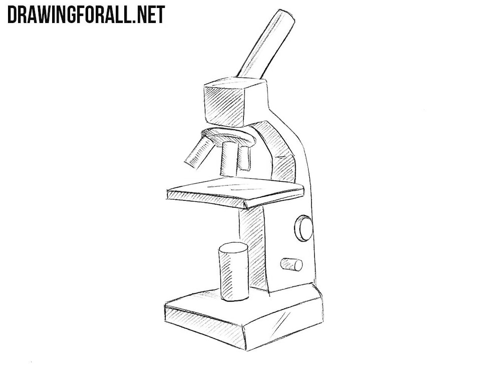
39 draw and label a binocular microscope
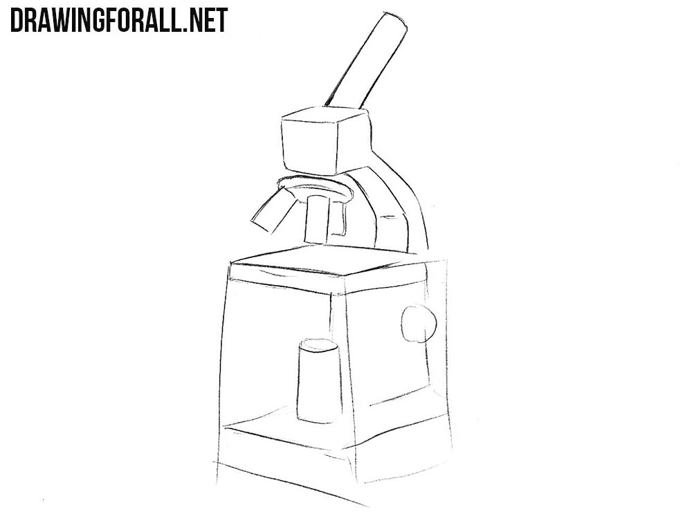
How to draw a microscope step by step beginners guide You are on the middle level then you can use a pencil,eraser, and paper. My recommendation uses always 2b pencil. Because It's a very light and soft pencil. Alyas first draw very lightly. step by step microscope drawing beginners Instaction. #Step 1 : #Step 2 : #Step 3 : #Step 4 : Label the microscope — Science Learning Hub Use this with the Microscope parts activity to help students identify and label the main parts of a microscope and then describe their functions. Drag and drop the text labels onto the microscope diagram. If you want to redo an answer, click on the box and the answer will go back to the top so you can move it to another box.
PDF Draw a microscope and label its parts Draw a binocular microscope and label its parts. Draw a compound microscope and label its parts. images. The lens is maintained near the object, a distance focal distance or less, And the eye is positioned near the lens on the other side. In standard microscopes, the goals are assembled so that when you switch between objectives, the sample ...

Draw and label a binocular microscope
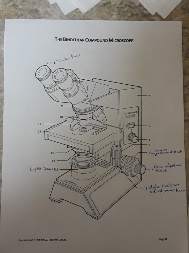
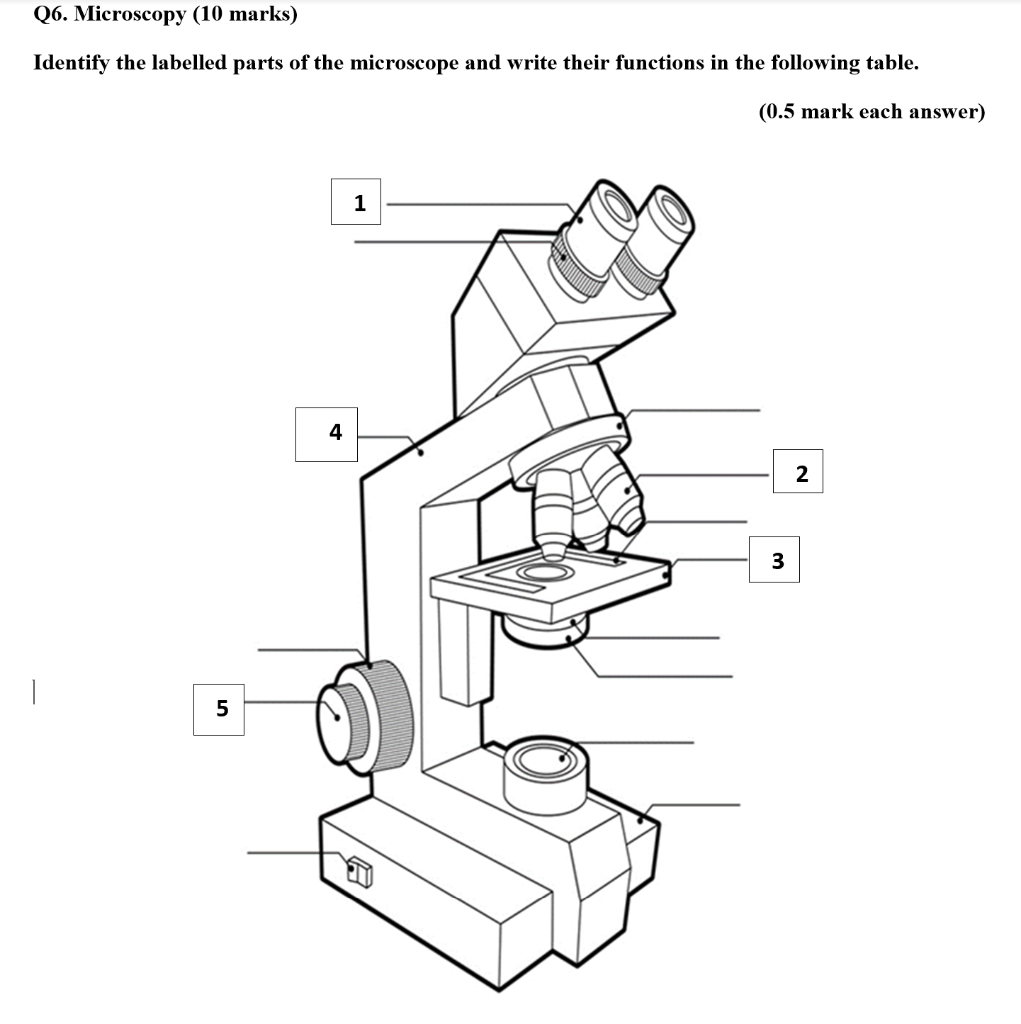
Solved Draw, label and mention function of parts of | Chegg.com Discuss how to use binocular microscope before, during and after. Question : Draw, label and mention function of parts of Binocular Microscope. This problem has been solved! Draw and Label Diagram Of Microscope - step by step | Adnan medicos Draw and Label Diagram Of Microscope - step by step | Adnan medicos | Parts of the Microscope with Labeling (also Free Printouts) Parts of the Microscope with Labeling (also Free Printouts) A microscope is one of the invaluable tools in the laboratory setting. It is used to observe things that cannot be seen by the naked eye. Table of Contents 1. Eyepiece 2. Body tube/Head 3. Turret/Nose piece 4. Objective lenses 5. Knobs (fine and coarse) 6. Stage and stage clips 7. Aperture
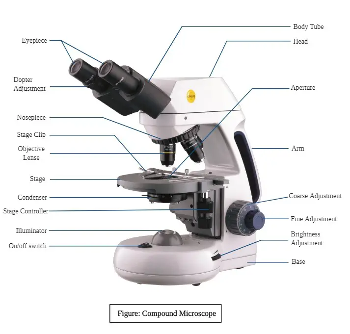
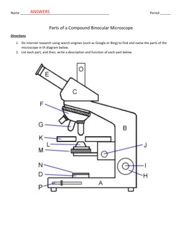
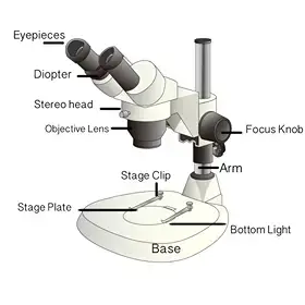
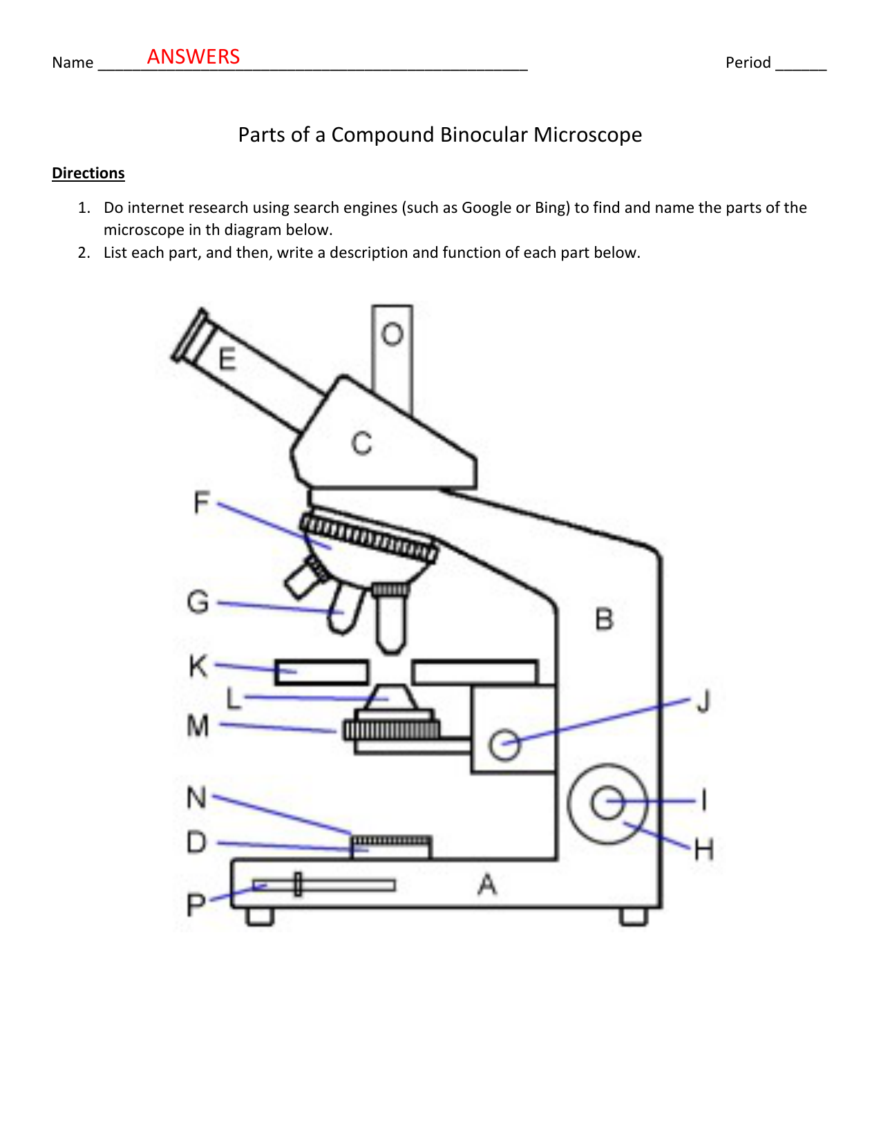
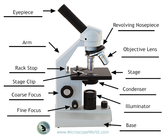
Draw and label a binocular microscope. Microscope Drawing Easy with Label - YouTube In this video I go over a microscope drawing that is easy with label. There is a blank copy at the end of the video to review on your own. A great way to s... Compound Microscope Parts, Functions, and Labeled Diagram Compound Microscope Definitions for Labels. Eyepiece (ocular lens) with or without Pointer: The part that is looked through at the top of the compound microscope. Eyepieces typically have a magnification between 5x & 30x. Monocular or Binocular Head: Structural support that holds & connects the eyepieces to the objective lenses. Question : hand draw a compound binocular light microscope and label ... hand draw a compound binocular light microscope and label its parts; Question: hand draw a compound binocular light microscope and label its parts. This problem has been solved! See the answer See the answer See the answer done loading. PDF Label parts of the Microscope: Answers Label parts of the Microscope: Answers Coarse Focus Fine Focus Eyepiece Arm Rack Stop Stage Clip . Created Date: 20150715115425Z ...
Microscope Drawing: How to Sketch Microscope Slides How to Draw Microscope Slides Organize and orient your field of view: To begin, draw a circle as large as possible with a pencil. An 8.5 x 11-inch piece of paper is good size for beginners. The circle represents what you see through the eyepiece of the microscope. Using thin lines, divide the circle into quarters in order to organize the picture. Labelled Diagram Of A Light Microscope | Products & Suppliers ... Knight Optical (UK) Ltd Custom Plane Mirrors for Microscopes.Plane mirrors also known as front surface mirrors or first surface mirrors are used widely within Microscope applications. As stock we hold a number of general purpose, l/1 and l/4 with a range of up to 6 types of coatings such as Enhanced Aluminium, Ali/SiO2 and Ali/Mgf2. Compound Microscope - Diagram (Parts labelled), Principle and Uses Also called as binocular microscope or compound light microscope, it is a remarkable magnification tool that employs a combination of lenses to magnify the image of a sample that is not visible to the naked eye. Compound microscopes find most use in cases where the magnification required is of the higher order (40 - 1000x). Microscope Parts, Function, & Labeled Diagram - slidingmotion Microscope parts labeled diagram gives us all the information about its parts and their position in the microscope. Microscope Parts Labeled Diagram The principle of the Microscope gives you an exact reason to use it. It works on the 3 principles. Magnification Resolving Power Numerical Aperture. Parts of Microscope Head Base Arm Eyepiece Lens
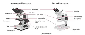
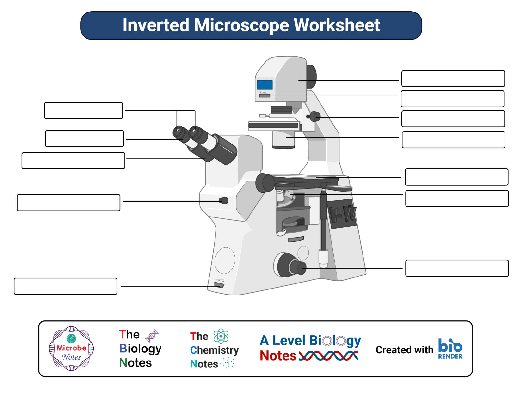
Parts of a microscope with functions and labeled diagram The eyepiece, also known as the ocular is the part used to look through the microscope. Its found at the top of the microscope. Its standard magnification is 10x with an optional eyepiece having magnifications from 5X - 30X. Objective Lens are the major lenses used for specimen visualization. They have a magnification power of 40x-100x. Microscope Labeling Diagram | Quizlet PGFry210. Unit 2 Lesson 5 - Punnett Squares and Pedigrees. 4 terms. PGFry210. Unit 2 Lesson 4 - Heredity. 9 terms. PGFry210. Upgrade to remove ads. Only $2.99/month. Compound Microscope Parts - Labeled Diagram and their Functions - Rs ... The eyepiece (or ocular lens) is the lens part at the top of a microscope that the viewer looks through. The standard eyepiece has a magnification of 10x. You may exchange with an optional eyepiece ranging from 5x - 30x. [In this figure] The structure inside an eyepiece. The current design of the eyepiece is no longer a single convex lens. Microscope, Microscope Parts, Labeled Diagram, and Functions Revolving Nosepiece or Turret: Turret is the part of the microscope that holds two or multiple objective lenses and helps to rotate objective lenses and also helps to easily change power. Objective Lenses: Three are 3 or 4 objective lenses on a microscope. The objective lenses almost always consist of 4x, 10x, 40x and 100x powers. The most common eyepiece lens is 10x and when it coupled with ...
Drawing Of A Microscope And Label - Warehouse of Ideas If you are drawing from a microscope, it is useful to state the combined magnification of the eyepiece plus objective lenses used when making the drawing, e.g. First to represent the microscope field of view draw a circle on the page this can be freehand or if you want to be precise use a compass. Source: What should a microscope drawing include.
Drawing Of A Binocular Microscope - Warehouse of Ideas Binocular microscope drawing with label color video biology online rules and of cells template. Source: paintingvalley.com. Surprising vaccine comic book style cartoon words. Its found at the top of the microscope. Source: For adapting the distance apart of. These microscopes can weigh approximately 130 pounds (60 kg) and can be quite large.
Microscope Diagram and Functions | Science fair projects, Microscope ... A Study of the Microscope and its Functions With a Labeled Diagram To better understand the structure and function of a microscope, we need to take a look at the labeled microscope diagrams of the compound and electron microscope. These diagrams clearly explain the functioning of the microscopes along with their respective parts. M mooketsi
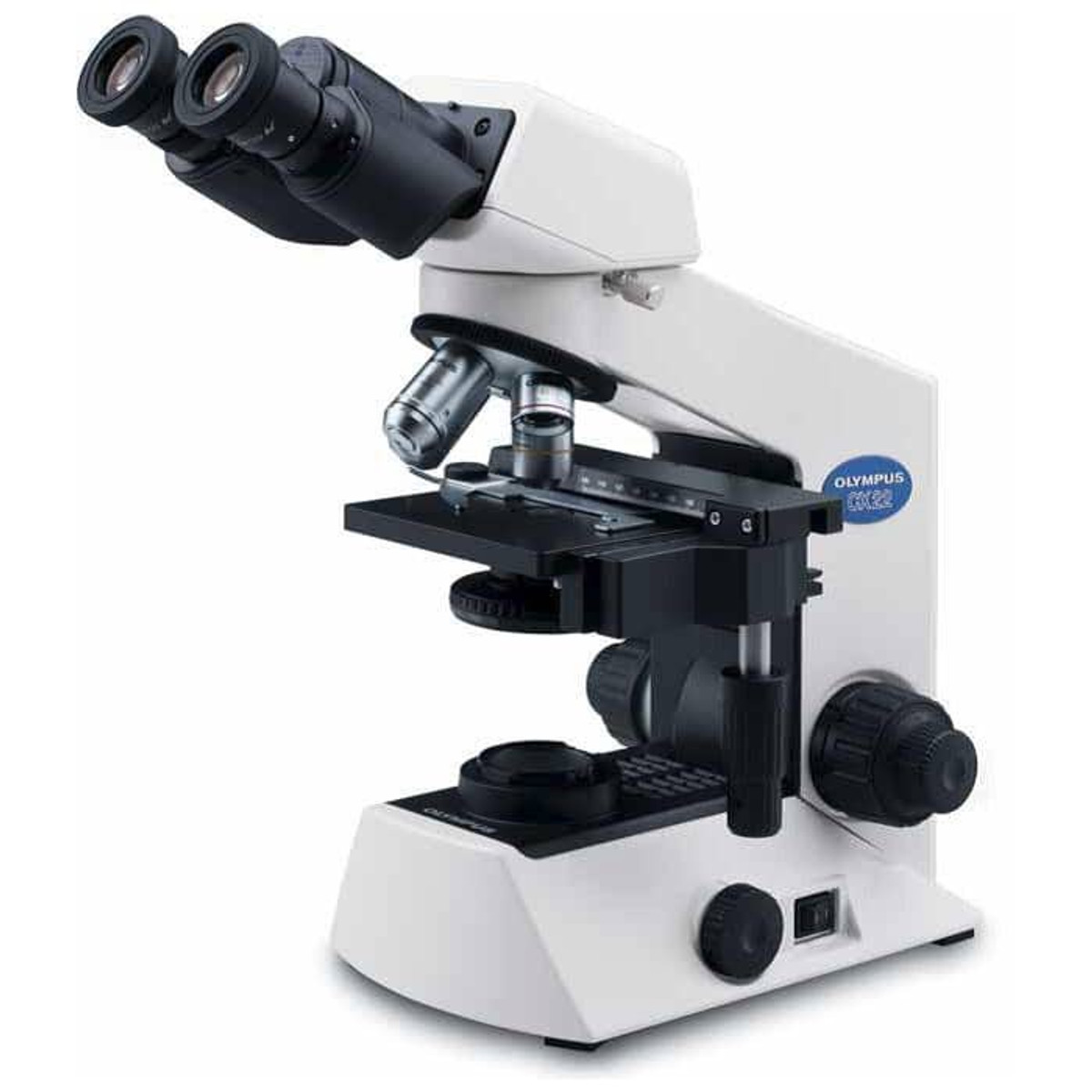
Microscope Parts & Functions - AmScope Base: A microscope is typically composed of a head or body and a base. The base is the support mechanism. Binocular Microscope: A microscope with a head that has two eyepiece lenses. Nowadays, binocular is typically used to refer to compound or high-power microscopes where the two eyepieces view through a single objective lens.
15+ How To Draw And Label A Microscope Background Draw and label a light microscope. Observe the skin cells under both low and high power of your microscope. Remember to subscribe to my channel and turn on notification. Microscope drawing and label at. Microscope drawing and label at getdrawings com free for. How to draw microscope slides. How to use a microscope.
How you can Label a Binocular Microscope - ScienceBriefss.com Identify the eyepieces at the top of the microscope. Binocular microscopes feature two separate eyepieces each with an ocular lens. Both the distance between the eyepieces and the focus of each eyepiece may be adjusted. Label the eyepieces. Find the nosepiece and objective lenses located beneath the eyepiece.
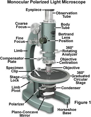
Microscope Parts and Functions Body tube (Head): The body tube connects the eyepiece to the objective lenses. Arm: The arm connects the body tube to the base of the microscope. Coarse adjustment: Brings the specimen into general focus. Fine adjustment: Fine tunes the focus and increases the detail of the specimen. Nosepiece: A rotating turret that houses the objective lenses ...
Compound Microscope- Definition, Labeled Diagram, Principle, Parts, Uses The optical microscope often referred to as the light microscope, is a type of microscope that uses visible light and a system of lenses to magnify images of small subjects. There are two basic types of optical microscopes: Simple microscopes. Compound microscopes. The term "compound" in compound microscopes refers to the microscope having ...
Diagrams of binocular microscope with labels? - Answers Binocular microscopes have a pair of eyepieces, each with two or more lenses. This allows the operator to use both eyes thus doing away with the eyestrain usually caused by a monocular microscope....
Labelled Diagram of Compound Microscope - Biology Discussion The below mentioned article provides a labelled diagram of compound microscope. Part # 1. The Stand: The stand is made up of a heavy foot which carries a curved inclinable limb or arm bearing the body tube. The foot is generally horse shoe-shaped structure (Fig. 2) which rests on table top or any other surface on which the microscope in kept.
Parts of the Microscope with Labeling (also Free Printouts) Parts of the Microscope with Labeling (also Free Printouts) A microscope is one of the invaluable tools in the laboratory setting. It is used to observe things that cannot be seen by the naked eye. Table of Contents 1. Eyepiece 2. Body tube/Head 3. Turret/Nose piece 4. Objective lenses 5. Knobs (fine and coarse) 6. Stage and stage clips 7. Aperture
Draw and Label Diagram Of Microscope - step by step | Adnan medicos Draw and Label Diagram Of Microscope - step by step | Adnan medicos |
Solved Draw, label and mention function of parts of | Chegg.com Discuss how to use binocular microscope before, during and after. Question : Draw, label and mention function of parts of Binocular Microscope. This problem has been solved!








Post a Comment for "39 draw and label a binocular microscope"