41 labeled diagram of nephron
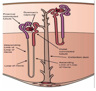
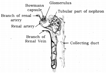
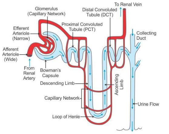
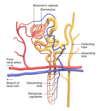
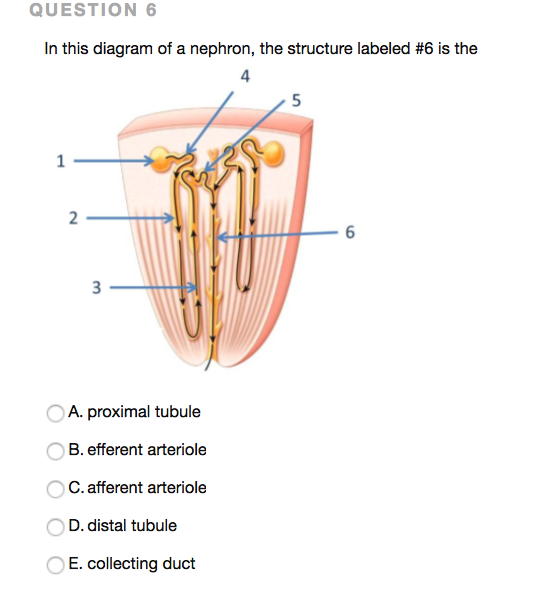
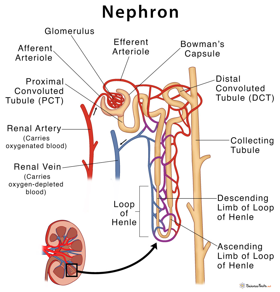
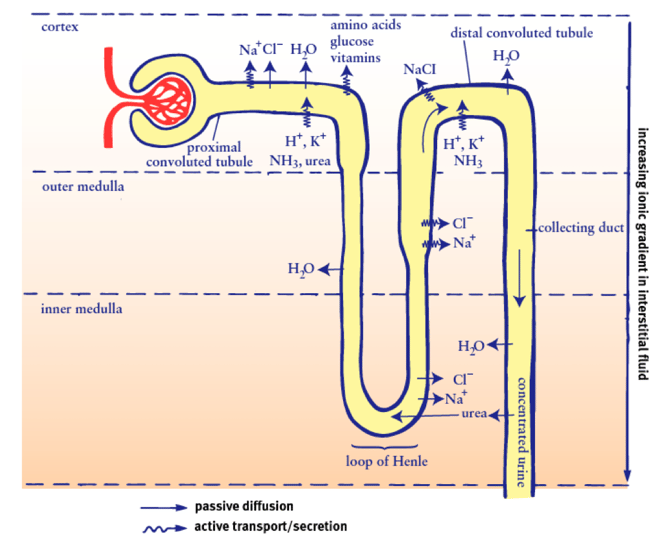
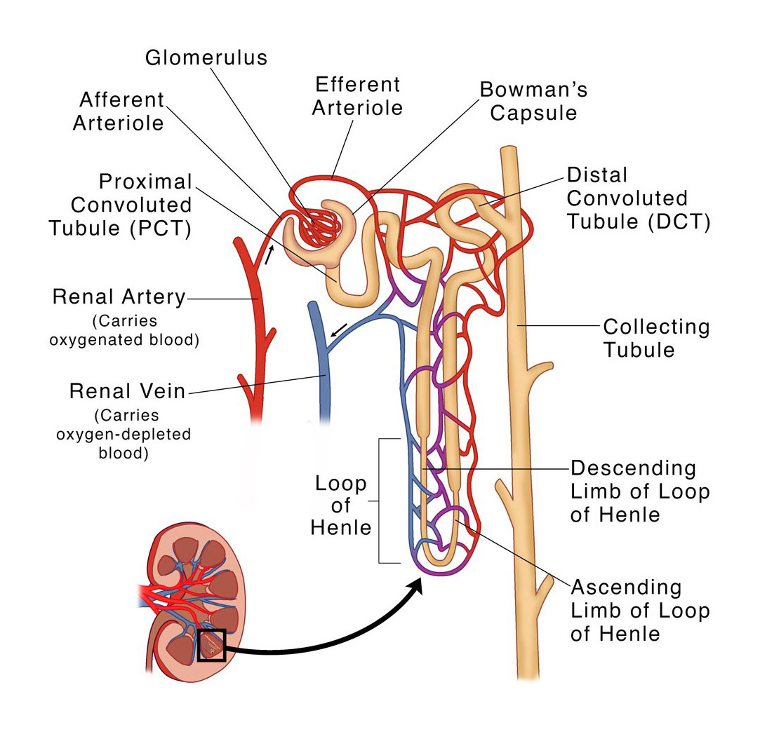
Breaking down nephron functioning into six easy steps! Here is a labeled diagram of a nephron followed by a general overview of the process: image sourced from Kaplan Step 1: Bowman's Capsule. Blood pressure in the small capillaries at one end of the nephron (called the glomerulus) pushes fluid into a sack called Bowman's capsule. Nephron- Definition, Structure, Parts, Functions, Diagram The basic structural and functional unit of the kidney, also known as the Nephron is a microscopic structure that is made up of both the renal tubule and renal corpuscle. The nephron is a Greek word that means kidney and one kidney can have millions of them. There are different stages of the development of the nephron.
Biology Questions and Answers Form 2 - High School Biology … Biology Questions and Answers Form 2; More than 5000 biology questions and answers to help you study biology. Online biology test questions and answers pdf, exam, quiz, test high school with answers. Biology syllabus. Biology questions and answers. Biology quiz with answers.

Labeled diagram of nephron
Nephron Structure, Function & Diagram - Study.com Nephrons are very minute tiny structures. The nephron of the kidney in mammals is a tube that is approximately 30-55 mm in length. The nephron has an inflated and closed tube. The end of this tube... Label a Nephron Quiz - PurposeGames.com This is an online quiz called Label a Nephron. There is a printable worksheet available for download here so you can take the quiz with pen and paper. From the quiz author. This is a nephron. Label it. This quiz has tags. Click on the tags below to find other quizzes on the same subject. biology. kidney. Nephron. simple diagram of nephron Nephron Diagram studylib.net. nephron. BIOLOGY: Structures Of The Nephron (Kidney Tubule) - Btec Biomedical Science With Rachael Verney . nephron kidney tubule biology structures structure name blank diagram. Structure Of The Nephron And Collecting System Quiz . Biology Nephron Diagram Labeled - Diagram Media
Labeled diagram of nephron. Nephron Labeled in 2022 | Science diagrams, Paper illustration, Labels in the musical labeled diagram, we have infraspinatus, teres major, triceps, gluteus medius, gluteus maximus, soleus, tibialis anterior, gastrocnemius, quadriceps, hamstrings, finger flexors, finger extensors, brachioradialis, external oblique, serratus anterior, latissimus dorsi, deltoid, trapezius, pectoralis major, biceps, abdominals, … Single-cell analysis identifies the interaction of altered renal ... 12.05.2022 · Fibrosis is the result of chronic inflammatory reactions induced by a variety of stimuli, including persistent infections, autoimmune reactions, allergic responses, chemical insults, radiation and ... Nephron - Wikipedia Fig.1) Schematic diagram of the nephron (yellow), relevant circulation (red/blue), and the four methods of altering the filtrate. The nephron is the functional unit of the kidney. [2] This means that each separate nephron is where the main work of the kidney is performed. A nephron is made of two parts: Diagram of nephron - Healthiack Diagram of nephron 193. Diagram of nephron 205. Diagram of nephron 206. Diagram of nephron 260. Diagram of nephron 269. Diagram of nephron 291. Diagram of nephron 394. Diagram of nephron 455. Diagram of nephron 727.
Annotated diagrams showing the nephrons up close - Mammoth Memory What a nephron looks like close up. This is what a nephron looks like - basically, it's a bendy tube (called a tubule) surrounded by tiny blood vessels. Substances are transferred between the blood vessels and the tube, to remove waste and adjust the balance of water and salts in the bloodstream. NOTE. What goes into the tubule is called ... Simple Diagram Of Nephron The nephron is the microscopic structural and functional unit of the kidney. It is composed of a Diagram (left) of a long juxtamedullary nephron and (right) of a short cortical nephron. . Proximal convoluted tubule (lies in cortex and lined by simple cuboidal epithelium with brush borders which help to increase the area of . Diagram of nephron Structure of the Kidney (With Diagram) | Organs | Human Physiology After reading this article you will learn about the structure of the kidney. This will also help you to draw the structure and diagram of kidney. The kidneys are two in number which are situated one on each side of the verteral column and in-front of the last ribs. They lie on the posterior abdominal wall. The right kidney is placed slightly ... Labeled Diagram of Nephron - YouTube Labeled Diagram of Nephron
Nephron diagram labeled - Healthiack This brief post displays Nephron diagram labeled … Please click on the picture (s) to view larger version. Feel free to search healthiack.com for more details on this very topic. Best viewed on 1280 x 768 px resolution in any modern browser. Nephron diagram labeled 800 Nephron diagram labeled 808 Nephron diagram labeled 815 CBSE Class 10 Answered - TopperLearning A kidney is composed of an enormous number of uriniferous tubules .They are also known as nephrons or renal tubules or kidney tubules. Nephrons are the structural and functional units of the kidney. Each kidney is formed of about 1 million nephrons. Nephrons are held together by a connective tissue. Structure of nephron: PDF Nephron Diagrams - Oak Park Unified School District / Overview Then, label the proximal convoluted tubule (PCT), distal convoluted tubule (DCT), loop of Henle, glomerular capsule, and glomerulus on the figure. Also label the collecting duct (not part of the nephron) on the figure. C Ltd* Figure 15-4 Nephron Definition - BYJUS A nephron is the basic structural and functional unit of the kidney. They are the microscopic structure composed of a renal corpuscle and a renal tubule. The word nephron is derived from the Greek word - nephros, meaning kidney. There are about millions of nephrons in each human kidney. Structure of Nephron
Stanford University UNK the , . of and in " a to was is ) ( for as on by he with 's that at from his it an were are which this also be has or : had first one their its new after but who not they have
Transport across Cell Membrane: 4 Ways | Biology ADVERTISEMENTS: Transport across cell membrane is classified into four ways: 1. Diffusion (Passive Transport) 2. Osmosis 3. Active Transport 4. Vesicular Transport. Cell membrane acts as a barrier to most, but not all molecules. Cell membranes are semi-permeable barrier separating the inner cellular environment from the outer cellular environment. Since the cell membrane is made […]
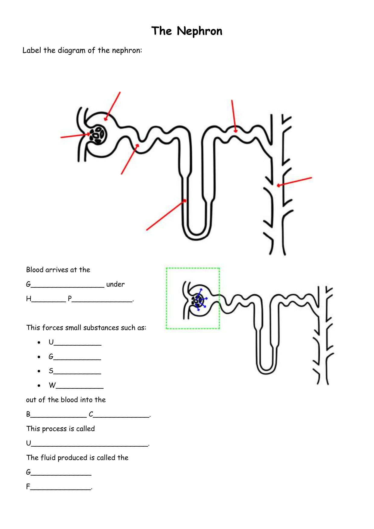
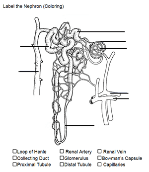
Color and Label the Nephron | Biology diagrams, Nursing school tips ... Practice labeling the nephron with this reinforcement activity. Students can also color the image to identify the major structures of the nephron: glomerulus, bowman's capsule, proximal and distal tubules, loop of henle, collecting duct and capillaries. This was designed to go with a larger unit on how the urinary system and kidneys help the body.
Nephron Diagram | How to draw labelled diagram of nephron | What is ... How to draw Nephron diagram. It is a labelled diagram of nephron. Specially for class 9,10,11,12. QUE = WHAT IS NEPHRON ? ANS = Nephron, functional unit of t...
Nephron Labeled | EdrawMax Template in the following nephron labeled diagram, we have shown renal vein (carries oxygen-depleted blood), renal artery (carries oxygenated blood), proximal convoluted tubule (pct), afferent arteriole, glomerulus, efferent arteriole, bowman's capsule, distal convoluted tubule (dct), collecting tubule, descending limb of loop of henle, ascending limb of …
Selina Solutions Concise Biology Class 10 Chapter 9 The ... - BYJUS (e) Name the two major steps involved in the formation of the fluid that passes down the part labeled ‘3’. Solution:-Ultrafiltration and selective reabsorption are the two major steps involved in the formation of the fluid that passes down part 3 ureter. 3. The following diagram represents a mammalian kidney tubule (nephron) and its blood ...
Andrew File System Retirement - Technology at MSU Andrew File System (AFS) ended service on January 1, 2021. AFS was a file system and sharing platform that allowed users to access and distribute stored content. AFS was available at afs.msu.edu an…
Draw a labelled diagram of nephron. - toppr.com Draw the well labelled diagram of human nephron and define haemodialysis. Medium. View solution > Draw a labelled diagram of a nephron. State the functions of glomerulitis, distal convoluted tubule and descending limb of Henle's loop. Easy. View solution > Draw a labelled diagram of the malpighian tubules transfer section.
Label This Diagram Of A Nephron. | Chegg.com | Biology diagrams ... Find Education Chart Biology Nephron Diagram Vector stock images in HD and millions of other royalty-free stock photos, illustrations and vectors in the Shutterstock collection. ... Human Nephron Model, labeled: Human Kidney Model, unlabeled: Human Kidney Model, labeled: Items not labeled in photo: (1) Renal Hilum - indented area where ureter ...
Kidney Diagram Nephron Illustrations & Vectors - Dreamstime Download 123 Kidney Diagram Nephron Stock Illustrations, Vectors & Clipart for FREE or amazingly low rates! New users enjoy 60% OFF. 194,155,077 stock photos online. ... Nephron structure labeled diagram. Human kidney medical diagram with a cross section of the inner organ. Anatomy of the kidney, vector illustration for medical education.Human ...
Nephron (Glomerulus and Tubule) Structure, Diagram, Functions The glomerulus is the first part of the nephron where fluid is filtered from the blood. It has two parts, namely the network of capillaries that transport the blood to the site (glomerular capillaries) and the enlarged head of the nephron which collects the filtered fluid (Bowman's capsule). This part of the nephron lies in the renal cortex.
Labeled Diagram of the Human Kidney - Bodytomy Labeled Diagram of the Human Kidney The human kidneys house millions of tiny filtration units called nephrons, which enable our body to retain the vital nutrients, and excrete the unwanted or excess molecules as well as metabolic wastes from the body. In addition, they also play an important role in maintaining the water balance of our body.
Draw a diagram of a nephron, and explain its structure. A nephron is the basic filtration unit of the kidney. It is a cluster of thin-walled blood capillaries. There are different parts of the nephron in which the formation of urine take place which is the main function of the kidney. Bowman's capsule and the glomerulus are together called as the glomerular apparatus.
Nephron - Definition, Function and Structure | Biology Dictionary A nephron is the basic unit of structure in the kidney. A nephron is used separate to water, ions and small molecules from the blood, filter out wastes and toxins, and return needed molecules to the blood. The nephron functions through ultrafiltration. Ultrafiltration occurs when blood pressure forces water and other small molecules through ...
Nephron Diagram | Quizlet Glomerulus A ball of capillaries surrounded by Bowman's capsule in the nephron and serving as the site of filtration in the vertebrate kidney. glomerular capusule (Bowman's Capsule) Urinary part of the the nephron that surrounds the glomerulus proximal convoluted tubule the first segment of a renal tubule efferent arteriole
draw the labelled diagram of a nephron - biology Q&A - BYJU'S draw the labelled diagram of a nephron - Get the answer to this question and access a vast question bank that is tailored for students.
Nephron labeling Diagram | Quizlet tiny ball of capillaries in the kidney, filters the blood. bowman's capsule. surrounds the glomerulus. proximal convoluted tubule. Site at which most of the tubular reabsorption occurs. distal convoluted tubule. Between the loop of Henle and the collecting duct; Selective reabsorption and secretion occur here. descending loop of henle.
Dopamine - Wikipedia Dopamine (DA, a contraction of 3,4-dihydroxyphenethylamine) is a neuromodulatory molecule that plays several important roles in cells. It is an organic chemical of the catecholamine and phenethylamine families. Dopamine constitutes about 80% of the catecholamine content in the brain. It is an amine synthesized by removing a carboxyl group from a molecule of its precursor …
nephron | Definition, Function, Structure, Diagram, & Facts The most advanced nephrons occur in the adult kidneys, or metanephros, of land vertebrates, such as reptiles, birds, and mammals. Each nephron in the mammalian kidney is a long tubule, or extremely fine tube, about 30-55 mm (1.2-2.2 inches) long. At one end this tube is closed, expanded, and folded into a double-walled cuplike structure.
simple diagram of nephron Nephron Diagram studylib.net. nephron. BIOLOGY: Structures Of The Nephron (Kidney Tubule) - Btec Biomedical Science With Rachael Verney . nephron kidney tubule biology structures structure name blank diagram. Structure Of The Nephron And Collecting System Quiz . Biology Nephron Diagram Labeled - Diagram Media
Label a Nephron Quiz - PurposeGames.com This is an online quiz called Label a Nephron. There is a printable worksheet available for download here so you can take the quiz with pen and paper. From the quiz author. This is a nephron. Label it. This quiz has tags. Click on the tags below to find other quizzes on the same subject. biology. kidney. Nephron.
Nephron Structure, Function & Diagram - Study.com Nephrons are very minute tiny structures. The nephron of the kidney in mammals is a tube that is approximately 30-55 mm in length. The nephron has an inflated and closed tube. The end of this tube...








:watermark(/images/watermark_5000_10percent.png,0,0,0):watermark(/images/logo_url.png,-10,-10,0):format(jpeg)/images/overview_image/1189/zTTH5bvyaS2CHjB48thWA_histology-nephron-vessels_english.jpg)






Post a Comment for "41 labeled diagram of nephron"