39 labelled diagram of microscope
Light microscopes - Cell structure - Edexcel - BBC Bitesize The magnification of a lens is shown by a multiplication sign followed by the amount the lens magnifies. So a lens magnifying ten times would be ×10. The total magnification of a microscope is:... (a) Draw the labelled ray diagram for the formation of image by a ... (a) Draw the labelled ray diagram for the formation of image by a compound microscope. Derive an expression for its total magnification (or magnifying power), when the final image is formed at the near point. (b) Why both objective and eyepiece of a compound microscope must have short focal lengths?
Labelled Diagram of Compound Microscope - Biology Discussion The below mentioned article provides a labelled diagram of compound microscope. Part # 1. The Stand: The stand is made up of a heavy foot which carries a curved inclinable limb or arm bearing the body tube. The foot is generally horse shoe-shaped structure (Fig. 2) which rests on table top or any other surface on which the microscope in kept.

Labelled diagram of microscope
Neuron under Microscope with Labeled Diagram - AnatomyLearner Neuron under microscope labelled diagram. Throughout this article, you got the different neurons labelled diagrams. Here, you will also find the diagrams of different neuron types under a microscope. The neuron diagram shows the different parts (axon, dendrites, and cell body) of the neurons. Draw a labelled diagram of an image formed by a compound microscope ... Draw a labelled diagram of an image formed by a compound microscope, with the image at least distance of distinct vision. Write any one expression for its magnifying power. Medium Solution Verified by Toppr Expression of magnifying power of a compound microscope is given by: m=− u ov o(1+ f eD) (a) Draw a labelled ray diagram of compound microscope, when final ... (a) Draw a labelled ray diagram of compound microscope, when final image forms at the least distance of distinct vision. (b) Why is its objective of short focal length and of short aperture, compared to its eyepiece? Explain. (c) The focal length of the objective is 4 cm while that of eyepiece is 10 cm. The object is placed at a distance of 6 cm from the objective lens.
Labelled diagram of microscope. Labeled Diagram Of A Stereo Microscope | Products & Suppliers ... Products/Services for Labeled Diagram Of A Stereo Microscope. Microscopes - (705 companies) Microscopes are instruments that produce magnified images of small objects Microscopes are instruments that produce a magnified image of a small object. They are used in many scientific and industrial applications. Microscope Parts and Functions With Labeled Diagram and ... Most specimens are mounted on slides, flat rectangles of thin glass. The specimen is placed on the glass and a cover slip is placed over the specimen. This allows the slide to be easily inserted or removed from the microscope. It also allows the specimen to be labeled, transported, and stored without damage. Parts of a Compound Microscope and Their Functions Compound microscope mechanical parts (Microscope Diagram: 2) include base or foot, pillar, arm, inclination joint, stage, clips, diaphragm, body tube, nose piece, coarse adjustment knob and fine adjustment knob.. Base: It's the horseshoe-shaped base structure of microscope.All of the other components of the compound microscope are supported by it. ... Parts of Microscope, Function, Names & Labeled Diagram ... Dec 24, 2021 · Microscope parts labeled diagram gives us all the information about its parts and their position in the microscope. Microscope Parts Labeled Diagram The principle of the Microscope gives you an exact reason to use it. It works on the 3 principles. Magnification Resolving Power Numerical Aperture. Parts of Microscope Head Base Arm Eyepiece Lens
Microscope, Microscope Parts, Labeled Diagram, and Functions Jan 19, 2022 · Microscope, Microscope Parts, Labeled Diagram, and Functions What is Microscope? A microscope is a laboratory instrument used to examine objects that are too small to be seen by the naked eye. It is derived from Ancient Greek words and composed of mikrós, “small” and skopeîn,”to look” or “see”. (a) Draw a labelled ray diagram of a compound microscope. (b) Derive an ... selected May 26, 2018 by Vikash Kumar Best answer (a) Labelled diagram of compound microscope. The objective lens form image A' B' near the first focal point ofeyepiece. (b) Angular magnification of objective lens m0 = linear magnification h'/h where L is the distance between second focal point of the objective and first focal point of eyepiece. Draw a neat labelled diagram of a compound microscope and explain its ... Description : It consists of two convex lenses separated by a distance. The lens near the object is called objective and the lens near the eye is called eye piece. The objective lens has small focal length and eye piece has of larger focal length. The distance of the object can be adjusted by means of a rack and pinion arrangement. A Study of the Microscope and its Functions With a Labeled Diagram To better understand the structure and function of a microscope, we need to take a look at the labeled microscope diagrams of the compound and electron microscope. These diagrams clearly explain the functioning of the microscopes along with their respective parts. Man's curiosity has led to great inventions. The microscope is one of them.
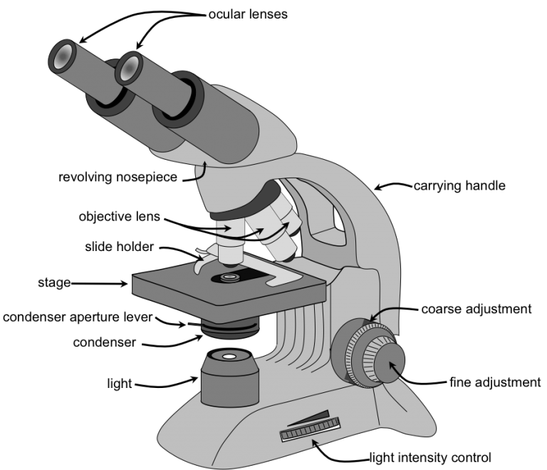
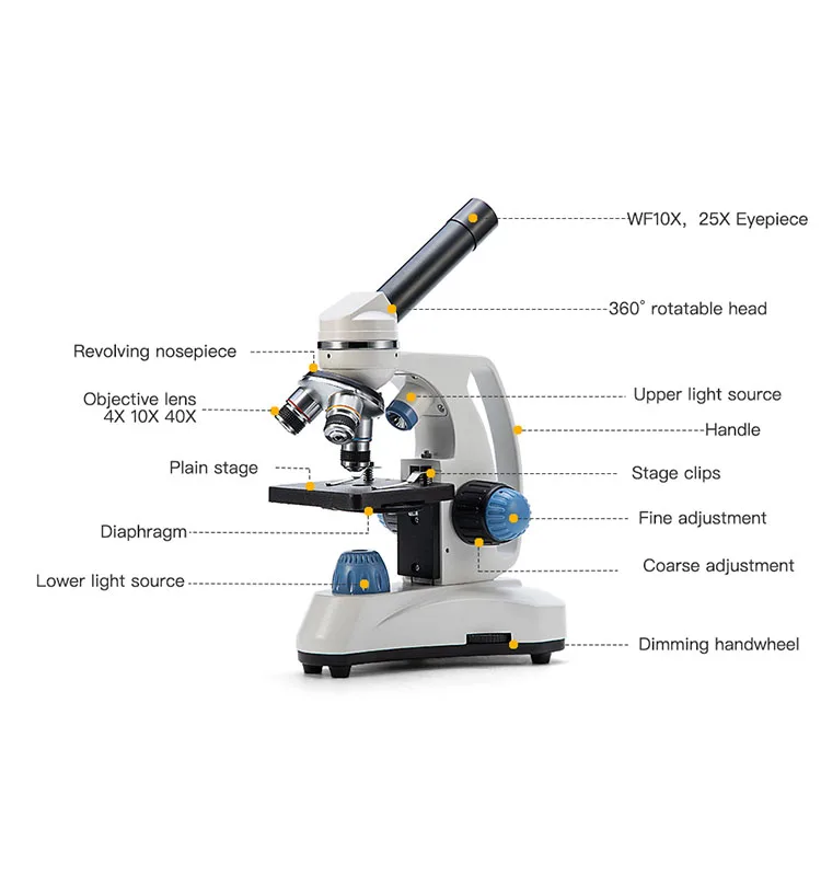
Parts of a microscope with functions and labeled diagram Apr 19, 2022 · Q. List down the 18 parts of a Microscope. 1. Ocular Lens (Eye Piece) 2. Diopter Adjustment 3. Head 4. Nose Piece 5. Objective Lens 6. Arm (Carrying Handle) 7. Mechanical Stage 8. Stage Clip 9. Aperture 10. Diaphragm 11. Condenser 12. Coarse Adjustment 13. Fine Adjustment 14. Illuminator (Light Source) 15. Stage Controls 16. Base 17. Labeled Microscope and Basics of Life Diagram | Quizlet PLAY. A microscope is an instrument widely to magnify and resolve the image of an object that is otherwise invisible to naked eye. For resolving the details of objects, which otherwise cannot be achieved by naked eye, a microscope is used. This set of flash cards will help the student to identify the different parts and function of the microscope. Compound Microscope Parts - Labeled Diagram and their Functions - Rs ... Labeled diagram of a compound microscope Major structural parts of a compound microscope There are three major structural parts of a compound microscope. The head includes the upper part of the microscope, which houses the most critical optical components, and the eyepiece tube of the microscope. Simple Microscope - Diagram (Parts labelled), Principle, Formula and Uses A simple microscope consists of Optical parts Mechanical parts Labeled Diagram of simple microscope parts Optical parts The optical parts of a simple microscope include Lens Mirror Eyepiece Lens A simple microscope uses biconvex lens to magnify the image of a specimen under focus.
Pin di Cells & Microscopes
Microscopes - Cell structure - AQA - GCSE Combined Science ... - BBC Late 1600s - Dutch scientist Antonie van Leeuwenhoek constructed a microscope with a single spherical lens. It magnified up to ×275. 1800s - the optical quality of lenses increased and the ...
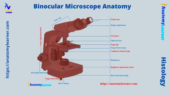
Binocular Microscope Anatomy - Parts and Functions with a ...
PDF Label parts of the Microscope Label parts of the Microscope: . Created Date: 20150715115425Z
Label a microscope - Teaching resources
Parts of Stereo Microscope (Dissecting microscope) - labeled diagram ... Labeled part diagram of a stereo microscope Major structural parts of a stereo microscope There are three major structural parts of a stereo microscope. The viewing Head includes the upper part of the microscope, which houses the most critical optical components, including the eyepiece, objective lens, and light source of the microscope.
Microscope Types (with labeled diagrams) and Functions
A Study of the Microscope and its Functions With a Labeled Diagram A Study of the Microscope and its Functions With a Labeled Diagram To better understand the structure and function of a microscope, we need to take a look at the labeled microscope diagrams of the compound and electron microscope. These diagrams clearly explain the functioning of the microscopes along with their respective parts. M mooketsi

5 Important Types of Microscopes used in Biology (With Diagram)
22 Parts Of a Microscope With Their Function And Labeled Diagram Parts Of a microscope. The main parts of a microscope that are easy to identify include: Head: The upper part of the microscope that houses the optical elements of the unit.; Base: The base is attached to a frame (arm) that is connected to the head of the device.The base of the microscope provides stability to the device and allows the user's hands to be free to manipulate other aspects of ...
Compound Microscope Parts, Functions, and Labeled Diagram ...
Compound Microscope Parts, Functions, and Labeled Diagram Nov 18, 2020 · Compound Microscope Parts, Functions, and Labeled Diagram Parts of a Compound Microscope Each part of the compound microscope serves its own unique function, with each being important to the function of the scope as a whole.

5 Important Types of Microscopes used in Biology (With Diagram)
Microscope labeled diagram - SlideShare Microscope labeled diagram 1. The Microscope Image courtesy of: Microscopehelp.com Basic rules to using the microscope 1. You should always carry a microscope with two hands, one on the arm and the other under the base. 2. You should always start on the lowest power objective lens and should always leave the microscope on the low power lens ...

Microscope Labeling Diagram | Quizlet
Label the microscope Diagram | Quizlet Start studying Label the microscope. Learn vocabulary, terms, and more with flashcards, games, and other study tools.

label microscope diagram | Charts | Microscope, Anatomy bones ...
Label the microscope — Science Learning Hub Use this interactive to identify and label the main parts of a microscope. Drag and drop the text labels onto the microscope diagram. eye piece lens coarse focus adjustment high-power objective diaphragm or iris base fine focus adjustment light source stage Download Exercise Tweet

Nikon Optiphot 66 Brightfield & Darkfield Reflected Light Microscope
Labeling the Parts of the Microscope Labeling the Parts of the Microscope This activity has been designed for use in homes and schools. Each microscope layout (both blank and the version with answers) are available as PDF downloads. You can view a more in-depth review of each part of the microscope here. Download the Label the Parts of the Microscope PDF printable version here.

How To Draw A Microscope, Step by Step, Drawing Guide, by ...
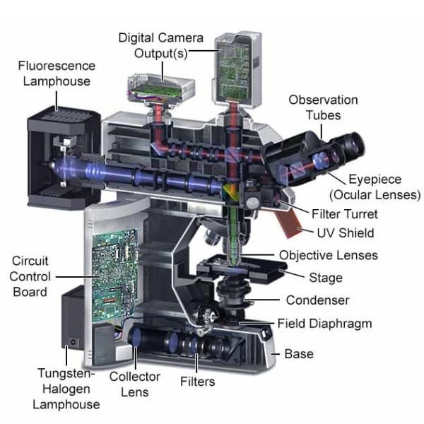
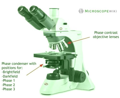
Microscope Types (with labeled diagrams) and Functions Dec 26, 2021 · Phase-contrast microscope labeled diagram Phase-contrast microscope functions: Its applications areas include In cases where the specimen is colorless and is very tiny In biology to conduct cellular level examination of microorganisms that can’t be visualized using the bright field microscopy Interference Microscope
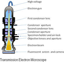
Electron microscope - Wikipedia
(a) Draw a labelled ray diagram of compound microscope, when final ... (a) Draw a labelled ray diagram of compound microscope, when final image forms at the least distance of distinct vision. (b) Why is its objective of short focal length and of short aperture, compared to its eyepiece? Explain. (c) The focal length of the objective is 4 cm while that of eyepiece is 10 cm. The object is placed at a distance of 6 cm from the objective lens.

How to Draw a Microscope and Label Its Parts
Draw a labelled diagram of an image formed by a compound microscope ... Draw a labelled diagram of an image formed by a compound microscope, with the image at least distance of distinct vision. Write any one expression for its magnifying power. Medium Solution Verified by Toppr Expression of magnifying power of a compound microscope is given by: m=− u ov o(1+ f eD)

Simple Microscope - Diagram (Parts labelled), Principle ...
Neuron under Microscope with Labeled Diagram - AnatomyLearner Neuron under microscope labelled diagram. Throughout this article, you got the different neurons labelled diagrams. Here, you will also find the diagrams of different neuron types under a microscope. The neuron diagram shows the different parts (axon, dendrites, and cell body) of the neurons.

Compound Microscope Parts – Labeled Diagram and their ...

Microscope diagram labeled | Clipart Panda - Free Clipart Images
مليمتر موردن محاسب condenser microscope function - poksipon.com
i) Draw a neat labelled ray diagram of a compound microscope ...

Microscope Diagram Labeled, Unlabeled and Blank | Parts of a ...
Fluorescence Microscopy - Explanation and Labelled Images ...
Swift Microscope Compound Microscopio 40x-1000x Monocular ...

Diagram of Compound Microscope

A labeled diagram of a microscope. MLT 101. :) | Teaching ...

File:Labelledmicroscope.gif - Wikimedia Commons
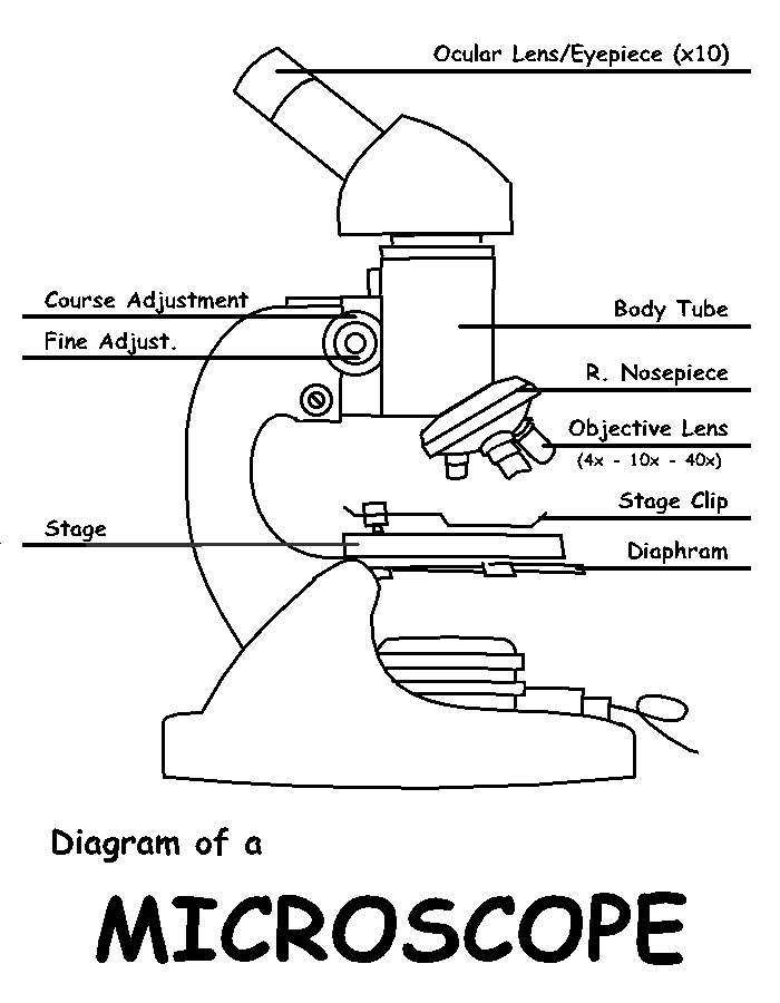
Diagram of a Microscope by ScienceDoodles on DeviantArt

Label Microscope Diagram - EnchantedLearning.com
Junior cert Labelling Microscope - Labelled diagram

microscope drawing with label - Clip Art Library

Microscope Label diagram Diagram | Quizlet

Microscopes: A Beginner's Guide

Compound Microscope – Diagram (Parts labelled), Principle and ...

Microscope With Labels clip art | Microscope parts ...
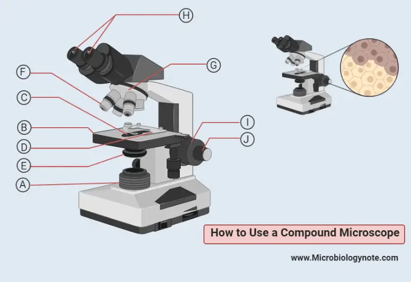
How to Use a Compound Microscope

PPT - Label the parts on your microscope picture. PowerPoint ...

Microscope Parts and Functions
Microscope - Teaching resources
Microscope Types (with labeled diagrams) and Functions
Microscope- Definition, Parts, Functions, Types, Diagram, Uses

Parts of a Microscope - SmartSchool Systems
Post a Comment for "39 labelled diagram of microscope"