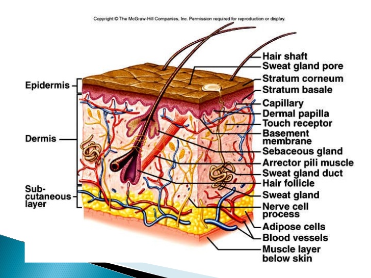
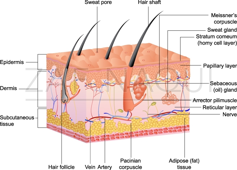
45 label the parts of the skin and subcutaneous tissue.
Subcutaneous Tissue Function and What Can Impact Its Health The middle layer of your skin contains sweat glands, lymphatic vessels, blood vessels, connective tissue, and hair follicles. Subcutaneous tissue. The deepest layer of skin is made of connective... What is Subcutaneous Tissue? - Medical News The subcutaneous tissue is composed of subcutaneous fat and various other types of cells. It is thickest in areas of the body such as the buttocks, palms, and soles of the feet. Subcutaneous fat is...
The Skin (Human Anatomy): Picture, Definition, Function, and Skin ... The dermis, beneath the epidermis, contains tough connective tissue, hair follicles, and sweat glands. The deeper subcutaneous tissue (hypodermis) is made of fat and connective tissue. The skin's...
Label the parts of the skin and subcutaneous tissue.
Skin Anatomy: The Layers of Skin and Their Functions Subcutaneous tissue is the innermost layer of the skin. It is mostly made up of fat, connective tissues, larger blood vessels, and nerves. 5 The majority of your body fat is stored in the subcutaneous layer. It not only insulates you against changing temperatures but protects your muscles and internal organs from impacts and falls. Anatomy and Physiology Homework Chapter 6 Flashcards - Quizlet The skin consists of two layers: a stratified squamous epithelium called the epidermis and a deeper connective tissue layer called the dermis. Below the dermis is another connective tissue layer, the hypodermis, which is not part of the skin. The skin is equipped with a variety of nerve endings that react to heat, cold, touch, texture, pressure, Layers of the Skin - Anatomy and Physiology The skin is composed of two main layers: the epidermis, made of closely packed epithelial cells, and the dermis, made of dense, irregular connective tissue that houses blood vessels, hair follicles, sweat glands, and other structures. Beneath the dermis lies the hypodermis, which is composed mainly of loose connective and fatty tissues.
Label the parts of the skin and subcutaneous tissue.. Label Skin Diagram Printout - EnchantedLearning.com Read the definitions, then label the skin anatomy diagram below. blood vessels - Tubes that carry blood as it circulates. Arteries bring oxygenated blood from the heart and lungs; veins return oxygen-depleted blood back to the heart and lungs. dermis - (also called the cutis) the layer of the skin just beneath the epidermis. Skin Anatomy - EnchantedLearning.com dermis - (also called the cutis) the layer of the skin just beneath the epidermis. epidermis - the outer layer of the skin. hair follicle - a tube-shaped sheath that surrounds the part of the hair that is under the skin. It is located in the epidermis and the dermis. Skin Cross-Section - Anatomy and Physiology - Innerbody Each layer is made of distinct tissues and performs distinct functions to support the body. A third layer of tissue under the skin, known as the hypodermis or subcutaneous layer, is not truly part of the skin itself but connects the skin loosely to the underlying muscles and bones that make up the deeper tissues of the body. The Skin: 7 Most Important Layers, Functions & Thickness The hypodermis is the deepest layer of skin situated below the dermis. It is also called the subcutaneous fascia or subcutaneous layer. It contains fat along with some structures like hair follicles, nerve endings, and blood vessels. The presence of fat helps insulate the body from heat and cold and serves as an energy storage area.
Genial Label The Skin Anatomy Diagram - Blogger Identify and label figures in Turtle Diarys interactive online game Skin Labeling. Label the skin anatomy diagram k. Label The Skin Anatomy Diagram - Fun for my own blog, on this occasion I will explain to you in connection with Label The Skin Anatomy Diagram.So, if you want to get great shots related to Label The Skin Anatomy Diagram, just click on the save icon to save the photo to your ... 5.1 Layers of the Skin - Anatomy & Physiology The hypodermis (also called the subcutaneous layer or superficial fascia) is a layer directly below the dermis and serves to connect the skin to the underlying fascia (fibrous tissue) surrounding the muscles. It is not strictly a part of the skin, although the border between the hypodermis and dermis can be difficult to distinguish. Function And Structure of Skin And Subcutaneous Tissue The epidermis is the thin outer layer of skin, the dermis is the thicker inner layer of skin. Beneath the dermis, lies a layer of loose connective tissue called subcutaneous tissue or the hypodermis deeper tissues including muscles, tendon, ligament, joint capsule and bone lie beneath the subcutaneous tissue layer. Subcutaneous Tissue: Composition, Function, Structure The outer part of the upper arm The middle part of the abdomen The front of the thigh The upper back The upper part of the buttocks The Effect of Age on Subcutaneous Tissue As you age, subcutaneous tissue starts to thin out. This weakened layer of insulation makes the body more sensitive to the cold because less tissue makes it harder to stay warm.
Chapter 6 Worksheet Flashcards - Quizlet Label the parts of the skin and subcutaneous tissue Label the layers of the skin top to bottom: - stratum corneum - stratum lucidum - stratum granulosum - stratum spinosum - stratum basale - dermis Label the cell types found in the skin Drag each label to the appropriate layer (A, B, or C) for each term or phrase. Skin: Cells, layers and histological features | Kenhub The epidermis is the uppermost layer of the skin. Going from deep to superficial, it consists of five layers; basal layer (stratum basale/germinativum) prickle cell layer (stratum spinosum) granular layer (stratum granulosum) clear layer (stratum lucidum) cornified layer (stratum corneum) To remember these layers, check out this mnemonics video: Integumentary System Parts Drawing / Label Skin Diagram Printout ... Integumentary System Parts Drawing / Label Skin Diagram Printout Enchantedlearning Com ... Structure and Functions of Skin - Anatomy, Diagram and ... - VEDANTU For example, the skin on the forearm which is on average 1.3 mm in the human male and 1.26 mm in the human female. The Structure of Human Skin Comprises Three Layers. The Three Layers of Skin Are. The outer layer of the skin: Epidermis. The part that consists of connective tissue: Dermis. The deepest layer of the skin: Subcutaneous tissue ...
Anatomy, Skin (Integument), Epidermis - StatPearls - NCBI Bookshelf It is made up of three layers, the epidermis, dermis, and the hypodermis, all three of which vary significantly in their anatomy and function. The skin's structure is made up of an intricate network which serves as the body's initial barrier against pathogens, UV light, and chemicals, and mechanical injury.
Hypodermis (Subcutaneous Tissue): Function & Structure The hypodermis is the bottommost layer of skin. What is the hypodermis? Your skin has three main layers: The hypodermis (subcutaneous tissue) is the innermost layer of skin in your body. The dermis is the middle layer. The epidermis is the outermost layer. Cleveland Clinic is a non-profit academic medical center.
Understanding The Different Layers Of Skin - SkinKraft Subcutaneous Tissue The subcutaneous tissue or hypodermis consists of well-vascularized, loose connective tissue and adipose tissue. The deeper tissues including muscle, tendon, ligament, joint capsule and bone that lie beneath the hypodermis. This layer maintains the temperature and acts like a cushion or shock absorber.
5.2 Accessory Structures of the Skin - Anatomy & Physiology Chapter Review. Accessory structures of the skin include hair, nails, sweat glands, and sebaceous glands. Hair is made of dead keratinized cells, and gets its color from melanin pigments. Nails, also made of dead keratinized cells, protect the extremities of our fingers and toes from mechanical damage. Sweat glands and sebaceous glands produce ...
Solved 6 Follow-up Label the parts of the skin and | Chegg.com Question: 6 Follow-up Label the parts of the skin and subcutaneous tissue Dermis Pressure receptor Sweatpores Cutaneous blood vessels Hairs Hypodermis Epidermis Sweatgland Reset Zoom
The subcutaneous layer: Anatomy, composition, and functions Subcutaneous tissue is the deepest skin layer that lies closest to the muscle. This layer has other names, including superficial fascia, hypodermis, subcutis, and tela subcutanea. The skin consists...
Solved Label the parts of the skin and subcutaneous tissue. | Chegg.com Question: Label the parts of the skin and subcutaneous tissue. 0.22 Cutaneous blood vessels Book Hypodermis Sweat pores Epidermis Hairs Dermis Lamellar corpuscle Sweat gland This problem has been solved! See the answer label the parts of the skin and subcutaneous tissue Show transcribed image text Expert Answer 98% (44 ratings) A …
Structure and Function of the Skin - MSD Manual Consumer Version The skin is the body's largest organ. It serves many important functions, including. Protecting the body against trauma. Regulating body temperature. Maintaining water and electrolyte balance. Sensing painful and pleasant stimuli. Participating in vitamin D synthesis. The skin keeps vital chemicals and nutrients in the body while providing a ...
[Solved] Please see an attachment for details | Course Hero HOW THE BODY WORKS Skin and Hair Directions: Print out and label the parts of the skin. WORD BANK epidermis subcutaneous tissue sweat gland dermis sebaceous gland hair HOW THE BODY WORKS The Nails Directions: Print out and label the parts of the nail. pale half-moon area of the nail where the nail meets the skin where nails begin under the skin WORD BANK nail root cuticle lunula EXTRAL Use the ...


Post a Comment for "45 label the parts of the skin and subcutaneous tissue."