40 correctly label the muscles of the leg
Muscles of the leg quizzes and labeled diagrams | Kenhub Leg muscles labeled Take a look at the leg muscles diagram below, where you see each muscle clearly labeled. Spend some time revising this diagram by connecting the name and location of each structure with what you've just learned in the video. The aim of this exercise is to improve your confidence in identifying different structures. PDF CHAPTER 10 Anatomy of the Muscular System Parallel muscles can vary in length, but long straplike muscles with parallel fascicles are perhaps most typical. The sartorius muscle of the leg is a good example. The rectus abdominis muscles, which run the length of the anterior abdominal wall, have parallel muscle fascicles that are "interrupted" by trans-verse intersections. 2.
Leg Muscles Anatomy, Function & Diagram | Body Maps Gastrocnemius (calf muscle): One of the large muscles of the leg, it connects to the heel. It flexes and extends the foot, ankle, and knee. Soleus: This muscle extends from the back of the knee to...

Correctly label the muscles of the leg
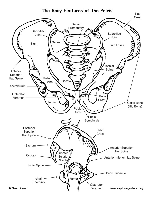
1.4 Anatomical Terminology - Anatomy & Physiology Figure 1.4.1 - Regions of the Human Body: The human body is shown in anatomical position in an (a) anterior view and a (b) posterior view. The regions of the body are labeled in boldface. A body that is lying down is described as either prone or supine. QUIZ 6 (CH11&12) Flashcards | Quizlet true. Correctly label the pectoral and brachial muscles. Correctly label the pectoral and brachial muscles. Correctly label the pectoral and brachial muscles. Label the anterior compartment muscles in this cross section through a forearm. Standing on your toes to reach a high shelf requires action of which muscle? Chapter 8/10 Flashcards | Quizlet Medial and lateral condyles of femur Ischial tuberosity Correctly label the muscles of the leg. Correctly label the bones and anatomical features of the male and female pelvis. The auditory ossicles include which of the following? Check all that apply. Hyoid Mastoid Match each label to its corresponding muscle of the quadriceps femoris.
Correctly label the muscles of the leg. Anatomical Body Landmark for Nursing - RNpedia Posterior Body Landmark. Calcaneal - heel of foot. Cephalic - head. Deltoid - curve of shoulder formed by large deltoid muscle. Femoral - thigh. Gluteal - buttock. Lumbar - area of back between ribs and hips. Occipital - posterior surface of head. Olecranal - posterior surface of elbow. Leg muscles | Human Anatomy Quiz - Quizizz Which muscle is #1? answer choices gastrocnemius sartorius gluteus medius gluteus maximus Question 9 120 seconds Q. Which muscle is #3? answer choices semimembranosus semitendinosus biceps femoris gastrocnemius Question 10 120 seconds Q. Which muscle is #4? answer choices gracilis semitendinosus semimembranosus biceps femoris Question 11 Connect Homework - Chapter 10 Flashcards | Quizlet Correctly label the muscles of the leg. Place a word or phrase from the left into each blank in a sentence on the right to make it correct. Correctly label the muscles of the anterior abdominal wall. What is the action of the orbicularis oris? Move each muscle to the action it is shown representing. Classify each muscle by its fascicle orientation. Muscles of the lower leg and foot | Human Anatomy and Physiology Lab ... The gastrocnemius muscle has two large bellies, called the medial head and the lateral head, and inserts into the calcaneus bone of the foot via its calcaneal tendon (also known as the Achilles tendon.) The soleus muscle is deep to the gastrocnemius, and the two muscles serve together as the calf of the leg.
Solved Correctly label the muscles of the leg. 33 Tibialis | Chegg.com Question: Correctly label the muscles of the leg. 33 Tibialis posterior 0.11 points Fibularis longus Gastrocnemius (cut) eBook Print References Fibularis brevis Flexor hallucis longus Calcaneal tendon (cut) Flexor digitorum longus Soleus (cut) Calcaneal tendon (cut) This problem has been solved! See the answer correctly label the muscles of the leg What Muscles Do Lunges Work? - SET FOR SET The main stabilizers are your hip abductors/adductors and the muscles of your lower legs. Note: In the forward lunge, movement should be slower in the eccentric phase as the hip, knee, and ankle joints move into flexion and faster in the concentric phase as power returns the forward leg to the standing position. Bones of the Lower Limb | Anatomy and Physiology | | Course Hero Like the upper limb, the lower limb is divided into three regions. The thigh is that portion of the lower limb located between the hip joint and knee joint.The leg is specifically the region between the knee joint and the ankle joint.Distal to the ankle is the foot.The lower limb contains 30 bones. These are the femur, patella, tibia, fibula, tarsal bones, metatarsal bones, and phalanges (see ... Key Muscle Locations and Movements - PT Direct For instance the quadriceps muscle group will extend the knee and flex the hip. The tables on the following pages detail the origin, insertion and action of some of the major muscles in the body. Use these tables in conjunction with the muscle charts on (in this same folder at ptdirect) to help you locate and understand how these muscles create ...
11.4 Identify the skeletal muscles and give their origins, insertions ... Editor's note: Replace figure with one that includes all muscles from table for example figure 10.7 from Marieb or 9.8 from Amerman. The orbicularis oris is a circular muscle that moves the lips, and the orbicularis oculi is a circular muscle that closes the eye. The occipitofrontalis muscle elevates the scalp and eyebrows. The muscle has a frontal belly and an occipital belly (near the ... Muscles of the Lower Limb - TeachMeAnatomy Muscles of the Leg. 3 Topics. Muscles of the Foot. View Article. Anatomy Video Lectures. START NOW FOR FREE. TeachMe Anatomy. Part of the TeachMe Series. The medical information on this site is provided as an information resource only, and is not to be used or relied on for any diagnostic or treatment purposes. This information is intended for ... Solved Correctly label the muscles of the leg. | Chegg.com Experts are tested by Chegg as specialists in their subject area. We review their content and use your feedback to keep the quality high. The given images belong to the muscle supply of the leg. Image 1 -flexor d …. View the full answer. Transcribed image text: Correctly label the muscles of the leg. Previous question Next question. Muscles of Leg- Labeling Flashcards - Quizlet Start studying Muscles of Leg- Labeling. Learn vocabulary, terms, and more with flashcards, games, and other study tools.
Angeline Ong Yoga World: Importance of Good Core Muscles
Solved Correctly label the muscles of the leg. Fibularis - Chegg Transcribed image text: Correctly label the muscles of the leg. Fibularis longus Fibularis brevis Tendon of plantaris Popliteus Flexor digitorum longus UUUUUUUUUU Plantaris Soleus Flexor hallucis longus Gastrocnemius (cut) Heads of gastrocnemius (cut) Reset Zoom Previous question Next question
30 Label The Muscles Of The Anterior Thigh. - Labels Information List
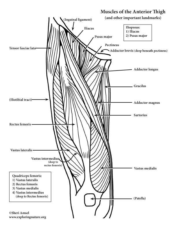
Chart of Major Muscles on the Front of the Body with Labels A muscle of the medial thigh that originates on the pubis. It inserts onto the linea aspera of the femur. It adducts, flexes, and rotates the thigh medially. It is controlled by the obturator nerve. It pulls the leg toward the body's midline (i.e. adduction) Biceps brachii An upper arm muscle composed of 2 parts, a long head and a short head.
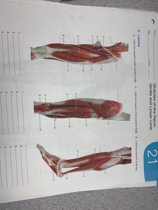
Solved: Muscles Of The Pelvic Girdle And Lower Limb 2 1 Na... | Chegg.com
Correctly label the muscles of the leg. Flashcards and Study Sets | Quizlet Learn Correctly label the muscles of the leg. with free interactive flashcards. Choose from 188 different sets of Correctly label the muscles of the leg. flashcards on Quizlet.
Patella Tracking Brace | Wraparound Hinged Knee Brace | BioSkin
PDF Muscle System Packet Key Part 2 - Gore's Anatomy & Physiology Muscles of the Hip, Thigh, and Leg 22. Identify the muscles described in Column A by choosing a response from Column B. Enter the correct letter in the answer blank. Select a different color for each muscle description provided with a color-coding circle, and use it to color the coding circles and corresponding muscles on Figure 6—9.

Anatomy And Physiology Archive | November 30, 2017 | Chegg.com
Muscles in the Lateral Compartment of the Leg - TeachMeAnatomy Revisions: 31. There are two muscles in the lateral compartment of the leg; the fibularis longus and brevis (also known as peroneal longus and brevis). The common function of the muscles is eversion - turning the sole of the foot outwards. They are both innervated by the superficial fibular nerve. In this article, we shall look at the anatomy ...
An Introduction to Skeletal System - The Bones and What They Do
Muscles Worksheet Flashcards & Practice Test | Quizlet Move each muscle to the action it is shown representing. Lateral Rotation: - trapezius (superior part) - serratus anterior Medial Rotation: - levator scapulae - rhomboideus major - rhomboideus minor Retraction: - rhomboideus major - rhomboideus minor - trapezius Move each muscle to the action it is shown representing. Elevation: - levator scapulae

FITNESS SOLUTIONS - ALL ABOUT FITNESS - EXERCISE - MUSCLE: 3/1/11 - 4/1/11
Solved Correctly label the muscles of the leg. Fibularis | Chegg.com Correctly label the muscles of the leg. Fibularis brevis Flexor hallucis longus Tendon of plantaris Flexor digitorum longus Heads of gastrocnemius (cut) Soleus ences Plantaris Fibularis longus Gastrocnemius (cut) Popliteus Reset Zoom Correctly label the muscles of the leg. Tibialis anterior Fibularis brevis Extensor digitorum longus Patella Soleus Patellar.

Chapter 8, Page 3 - HistologyOLM
Free Science Flashcards about ANP1040 Exam 3 - StudyStack Correctly label the posterior muscles of the thigh. Semitendinosus, Gluteus maximus, Biceps femoris, Semimembranosus, Gracillis: Correctly label the muscles of the leg. Calcaneus, Gastrocnemius (Medial Head), Grastrocnemius (Lateral Head), Tendon of Gastrocnemius: Correctly label the following parts of a skeletal muscle fiber.
Post a Comment for "40 correctly label the muscles of the leg"