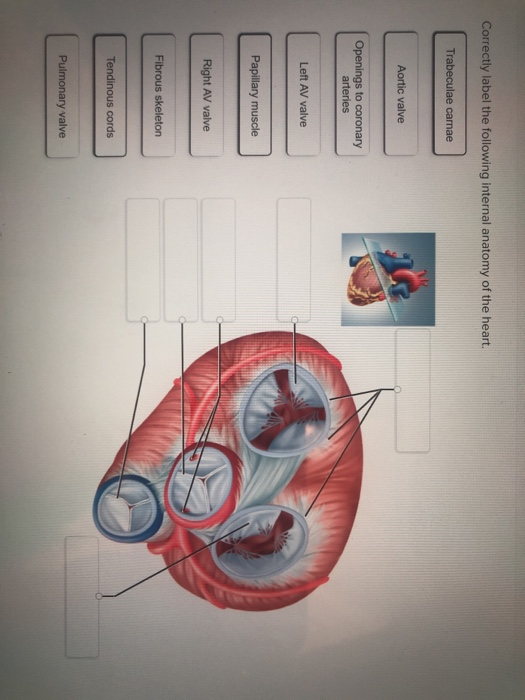
44 correctly label the following internal anatomy of the heart.
[Solved] Check my work Correctly label the followiting Internal anatomy ... e correcty label the following internal anatomy of the heart left atrium interventricular septum papillary muscles right ventricle opening of superior left ventricle vena cava fossa ovalis right atrium right atrium pectinate tricuspid valve muscles right ventricle rabeculae camae papillary muscle o download attachments: attachment1.png skip … Ch. 19 Circulatory System- heart Flashcards | Quizlet Correctly label the following internal anatomy of the heart. Drag each label to the location of each structure described. Explanation The heart functions to first pump deoxygenated blood returning from the body to the lungs in order to release carbon dioxide and reoxygenate the blood.
11 A&p ideas | anatomy and physiology, physiology, human anatomy and ... Feb 13, 2019 - Explore Imissthe Oldme's board "a&p" on Pinterest. See more ideas about anatomy and physiology, physiology, human anatomy and physiology.
Correctly label the following internal anatomy of the heart.
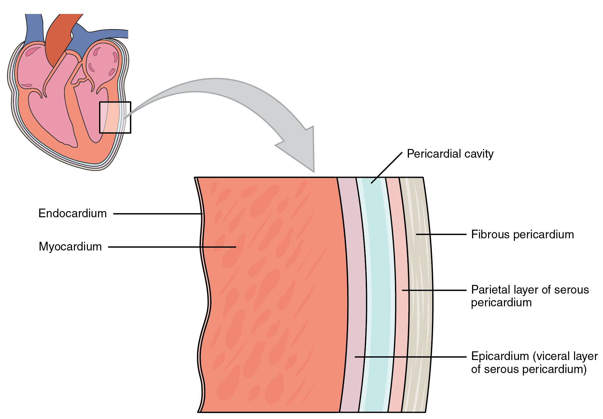
Chapter 11 Cardiovascular System Answers yourself before your exam. Start studying the Chapter 20-Cardiovascular System flashcards containing study terms like Correctly label the following internal anatomy of the heart., Correctly label... Answered: Label name correctly | bartleby Human Anatomy & Physiology (11th Edition) 11th Edition. ISBN: 9780134580999. Author: Elaine N. Marieb, Katja N. Hoehn. Publisher: PEARSON. expand_less. 1 The Human Body: An Orientation 2 Chemistry Comes Alive 3 Cells: The Living Units 4 Tissue: The Living Fabric 5 The Integumentary System 6 Bones And Skeletal Tissues 7 The Skeleton 8 Joints 9 ... Layers of the heart: Epicardium, myocardium, endocardium | Kenhub The myocardium is functionally the main constituent of the heart and the thickest layer of all three heart layers. It is a muscle layer that enables heart contractions. Histologically, the myocardium is comprised of cardiomyocytes.Cardiomyocytes have a single nucleus in the center of the cell, which helps to distinguish them from skeletal muscle cells that have multiple nuclei dispersed in the ...
Correctly label the following internal anatomy of the heart.. How the Heart Works: Diagram, Anatomy, Blood Flow The heart is about the size of a closed fist, weighs about 10.5 ounces, and is somewhat cone-shaped. It is covered by a sack termed the pericardium or pericardial sack. The normal heart anatomy consists of a four-chambered, hollow organ. It is divided into the left and right sides by a muscular wall called the septum. Heart Anatomy: Labeled Diagram, Structures, Blood Flow ... - EZmed Image: Use the 2x2 table to label the 4 chambers of the heart, including the right atrium, right ventricle, left atrium, and left ventricle. Tricuspid Valve and Mitral Valve Now that we have a good understanding of the 4 chambers of the heart, let's move on to the 4 main valves. Structure of the Heart | SEER Training Structure of the Heart. The human heart is a four-chambered muscular organ, shaped and sized roughly like a man's closed fist with two-thirds of the mass to the left of midline. The heart is enclosed in a pericardial sac that is lined with the parietal layers of a serous membrane. The visceral layer of the serous membrane forms the epicardium. Chapter 20-Cardiovascular System Flashcards - Quizlet Correctly label the following internal anatomy of the heart. b Place the labels in order denoting the flow of oxygenated blood through the heart beginning with the vessels that bring blood back to the heart from the lungs. Correctly label the following coronary blood vessels of the heart.
Correctly label the following internal anatomy of the heart. Fossa ... Correctly label the following parts of the internal anatomy of the heart. Place your cursor over the boxes for more information papillary muscles bicuspid valve right atrium septum pulmonary semilunar valve eft atrium chordae tendineae pulmonary... Label the heart - Science Learning Hub In this interactive, you can label parts of the human heart. Drag and drop the text labels onto the boxes next to the diagram. Selecting or hovering over a box will highlight each area in the diagram. Pulmonary vein Right atrium Semilunar valve Left ventricle Vena cava Right ventricle Pulmonary artery Aorta Left atrium Download Exercise Tweet Solved Correctly label the following parts of the internal - Chegg Correctly label the following parts of the internal anatomy of the heart. Right pulmonary veins Aorta Left pulmonary veins Pulmonary semilunar Left ventricle Right ventricle Bicuspid valve Tricuspid valve Right pulmonary artery Septum Pulmonary trunk Right atrium Left pulmonary artery Inferior vena cava Left atrium Superior vena cava Reset Zoom Anatomy and Function of the Heart's Electrical System ... In the simplest terms, the heart is a pump made up of muscle tissue. Like all muscle, the heart needs a source of energy and oxygen to function. The heart's pumping action is regulated by an electrical conduction system that coordinates the contraction of the various chambers of the heart.
Quiz 4 - Quiz4 1. Award: 0 out of 1.00 point Classify the following ... Award: 1 out of 1.00 point Correctly label the following parts of the internal anatomy of the heart. References Labeling Section: 05.03 3. Award: 1 out of 1.00 point Using the image as a guide, sequence the following descriptions of the flow of blood through the human heart. Correctly label the following internal anatomy of the heart. Right ... Right pulmonary artery Pulmonary trunk. Correctly label the following internal anatomy of the heart. Right pulmonary artery Pulmonary trunk Right pulmonary veins Pulmonary valve Right ventricle Aorta Left pulmonary veins Left pulmonary artery Right atrium Apr 01 2022 05:38 PM Expert's Answer Solution.pdf Next Previous Heart Labeling Quiz: How Much You Know About Heart Labeling? The human heart is a vital organ for every human. The more healthy your heart is, the longer the chances you have of surviving, so you better take care of it. Take the following quiz to know how much you know about your heart. Questions and Answers 1. What is #1? 2. What is #2? 3. What is #3? 4. What is #4? (HINT: it's a valve) 5. What is #5? Internal Structure of the Heart | Contemporary Health Issues The valves between the atria and ventricles are known generically as the tricuspid (right side)and the bicuspid (left side) va lve. The valves at the openings that lead to the pulmonary trunk and aorta are known generically as the pulmonary and the aortic valve. Structures of the Heart
19.1 Heart Anatomy – Anatomy and Physiology
The Anatomy of the Heart, Its Structures, and Functions The heart is the organ that helps supply blood and oxygen to all parts of the body. It is divided by a partition (or septum) into two halves. The halves are, in turn, divided into four chambers. The heart is situated within the chest cavity and surrounded by a fluid-filled sac called the pericardium. This amazing muscle produces electrical ...
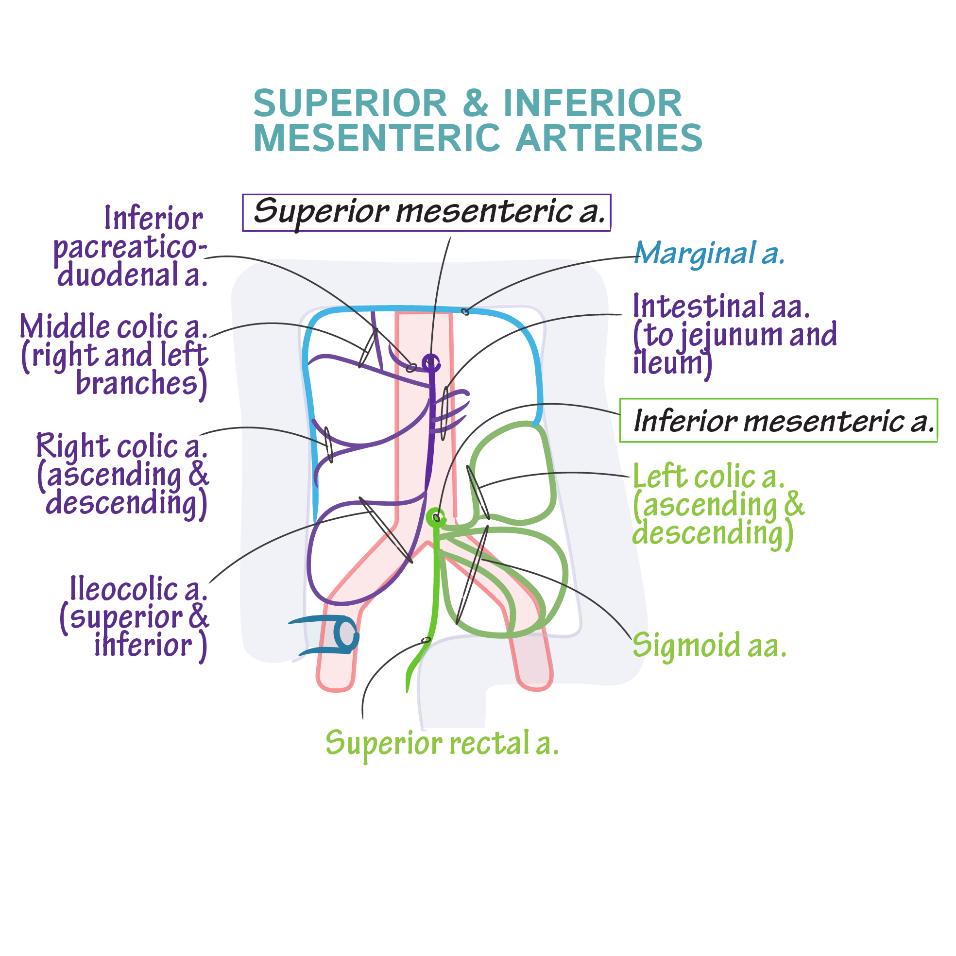
Superior Inferior Anatomy - Anatomy Drawing Diagram
Answered: Label name correctly please answer the… | bartleby Label name correctly please answer the following questions. Transcribed Image Text: Identify the structures of the placenta and umbilical cord on a model or chart of the placenta. Sinusoid- maternal blood Chorion Umbilical vein Umbilical arteries 1. 2 Intervillous space (containing maternal blood) Maternal blood Development of chorionic villi ...

ANATOMY OF HEART (PART-III) - YouTube
Cardiovascular System Heart Chapter 20 - hex.arista.com Correctly label the following internal anatomy of the heart. b, Place the labels in order denoting the flow of oxygenated blood through the heart beginning with Simplified relationship of the serous pericardium to the heart. A B. 326 CHAPTER 11 The CardiovascularSystem The Heart Ensures Continual, 24/7 Nutrient Delivery 327. The Heart
Heart Diagram with Blood Flow - peshsexam2
Heart Anatomy | Anatomy and Physiology I - Lumen Learning Relate the structure of the heart to its function as a pump. Compare systemic circulation to pulmonary circulation. Identify the veins and arteries of the coronary circulation system. Trace the pathway of oxygenated and deoxygenated blood thorough the chambers of the heart. The vital importance of the heart is obvious.

34 Correctly Label The Following Anatomical Features Of The Lymph Node ...
Human Heart (Anatomy): Diagram, Function, Chambers, Location in ... - WebMD The heart is a muscular organ about the size of a fist, located just behind and slightly left of the breastbone. The heart pumps blood through the network of arteries and veins called the...
Solved: Correctly Label The Following Internal Anatomy Of ... | Chegg.com
The Heart - Science Quiz - Seterra This science quiz game will help you identify the parts of the human heart with ease. Blood comes in through veins and exists via arteries—to control the direction of the flow, the heart has four sets of valves. The heart is an amazing machine with a lot of moving parts—let this quiz game help you find your way around this most vital of organs.
Post a Comment for "44 correctly label the following internal anatomy of the heart."